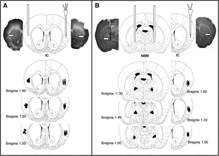
Figure 1.
Coronal section diagrams and representative photomicrographs of cannulae and probe placement in the IC and NBM. A, Groups with one stainless steel cannula in the left IC and one dual probe (injector and microdialysis probe) in the right IC. B, Groups with bilateral NBM injection cannulae and one cannula/microdialysis probe in the right IC. Arrows (above) and dots (below) show locations of stainless steel cannulae. Lines show microdialysis probes (modified from Paxinos and Watson, 1998).

