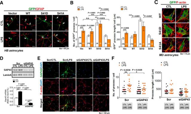
Figure 5.
GAP43 mediated LPS-induced astrocyte process arborization. A, Immunofluorescent confocal images of GFP (green), GFAP (red), and DAPI (blue) in primary rat HB astrocytes transfected with GFP-tagged WT-GAP43, S41D, or S41A GAP43 mutants for 24 h followed by 24 h of LPS (50 ng/ml) treatment. B, Quantification of the process number (left) and length (right) per cell in A. n.s., No significant difference. The number of microscopic images counted in each group was as indicated in the bar graph. C, Immunofluorescent confocal images of GFP (green) and F-actin (red) in primary rat MD astrocytes transfected with GFP-tagged WT-GAP43, S41D, or S41A GAP43 mutants followed by LPS treatment of the same condition as in A. Note that cellular hypertrophy, not astrocytic process formation, was observed in the S41D-GAP43-transfected or LPS-treated WT-GAP43-transfected astrocytes. D, Western blotting and qRT-PCR (bottom) of GAP43 expression in HB astrocytes transfected with siGAP43 or scrambled RNA (Scr) followed by vehicle (CTL) or LPS (50 ng/ml) treatment for 24 h to indicate the efficiency of the knockdown of GAP43. n = 3. E, Confocal immunofluorescence of GAP43 and F-actin (phalloidin labeling in red) indicated that siGAP43 transfection attenuated the LPS-induced GAP43 and affected astrocyte process arborization. F, Quantification of average astrocyte process number (left) and length (right) per cell obtained from E. The number of microscopic images counted in each group was as indicated below the graph.

