Figure 9.

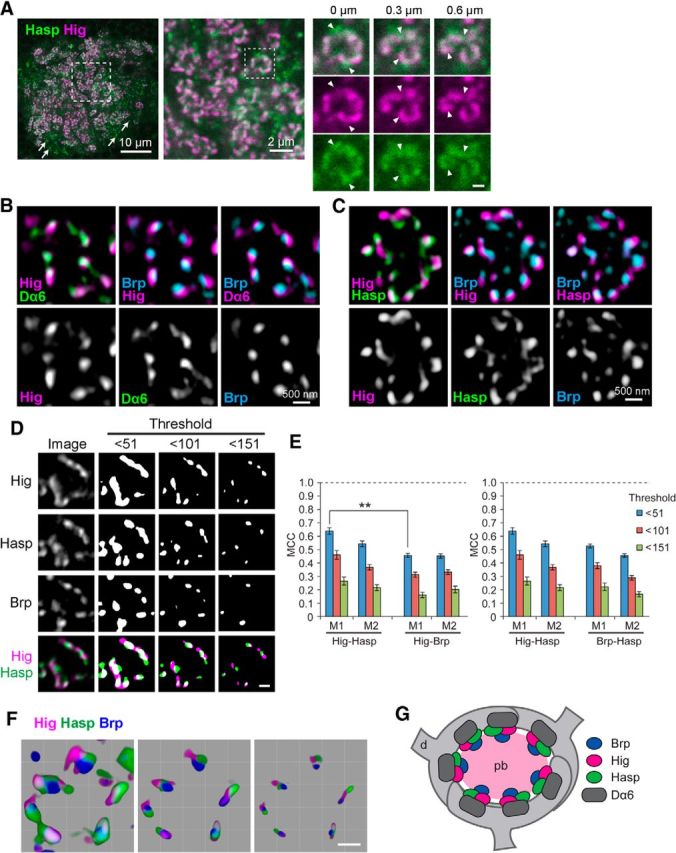
Hig and Hasp form matrix compartments in synaptic clefts. A, Confocal images of MB calyx. Both Hasp (green) and Hig (magenta) are closely associated in microglomeruli, but only Hasp is visible in nonmicroglomerular synapses (arrows); Hig is detectable only in overexposures. Dotted square in leftmost panel is magnified in the middle panel, and the region of a single microglomerulus (dotted square in the middle panel) is enlarged and shown as three sets of serial optical sections with Z intervals of 300 nm. Scale bar: serial image, 500 nm. Hasp does not completely overlap with Hig, as indicated by triangles. B, C, High-resolution images of single microglomeruli obtained by structured illumination microscopy. Optical sections triply immunolabeled for Hig, Dα6, and Brp (B) or Hig, Hasp, and Brp (C) are shown with two colors for each image. Hig was closely associated with presynaptic Brp and postsynaptic Dα6 (B). Hig and Hasp were located in close proximity but occupy distinct areas in the same synaptic cleft associated with Brp puncta (C). D, Binary images of a single microglomerulus triply labeled for Hig, Hasp, and Brp. The original image is shown in the leftmost column. Merged figures are shown at the bottom. Nonuniform distribution of the three proteins becomes clearer as the signal threshold is set higher. Binary images were processed after removal of signals with intensities lower than the threshold indicated at the top of each column. E, MCCs. As the threshold is increased, a similar reduction in MCC is observed for Hig-Hasp, Hig-Brp, and Hasp-Brp. Error bars indicate ± SEM. **p < 0.01, relative to M1 of Hig-Hasp with the same threshold (Mann–Whitney U test). F, Three-dimensional reconstruction images of a single microglomerulus. The threshold of image was arbitrarily changed, with the lowest at the left and the highest at the right. Scale bars: D, F, 500 nm. G, Schematic model for the distribution of Brp, Hig, Hasp, and Dα6 in a microglomerulus. Hig and Hasp localize to synaptic clefts. pb, Presynaptic bouton of olfactory projection neuron; d, dendrite of MB Kenyon cell.
