
Figure 1.
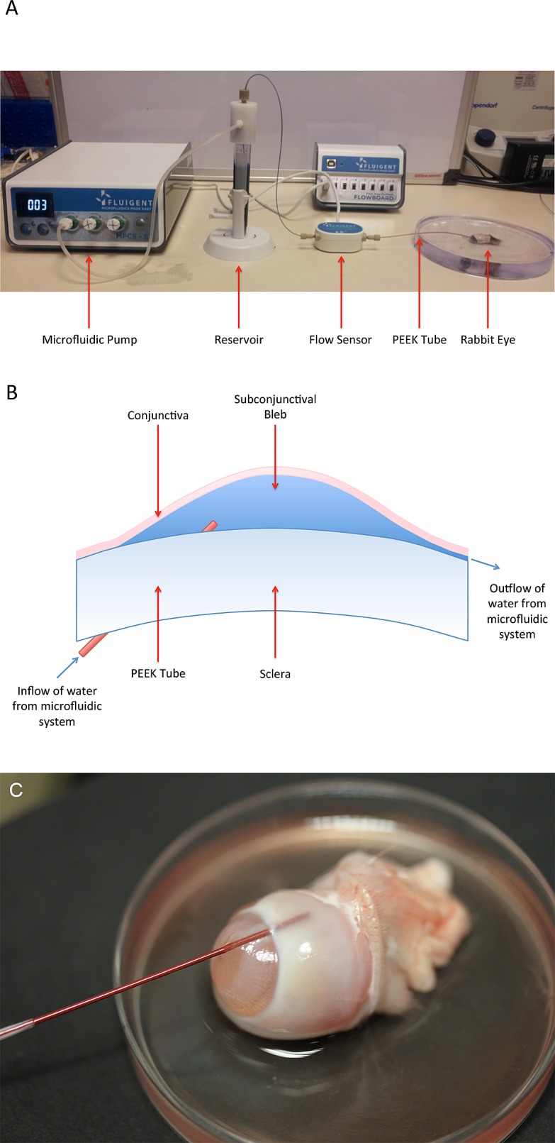
(A) Microfluidic setup demonstrating the pump pushing water from the reservoir through the flow sensor to the eye. (B) Schematic of fluid flow from the microfluidic setup into the subconjunctival space of the rabbit eye. (C) Close-up photograph demonstrating location of tube entering the eye through the cornea, into the anterior chamber angle and out into the subconjunctival space.