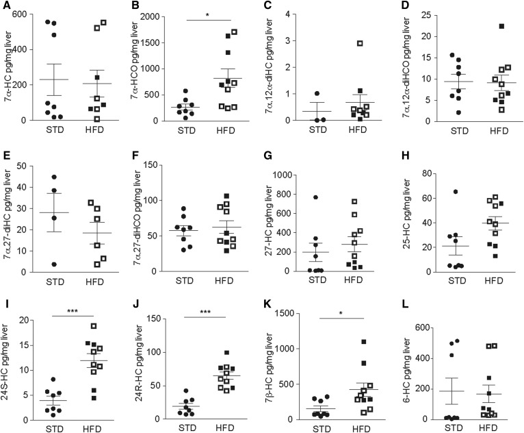
Fig. 6.
Liver oxysterol levels in a murine model of NAFLD/NASH. Eight-week-old male C57BL/6 mice were fed an HFD or STD for 20 weeks. Levels of the indicated oxysterol in the liver tissue of HFD (black squares: NASH; white squares: NAFLD) and STD controls were measured by LC-MS. Statistical analysis: Mann-Whitney U test (nSTD = 8, ≥3 valid data points; nHFD = 10, ≥5 valid data points). ***P < 0.001, **P < 0.01, and *P < 0.05.

