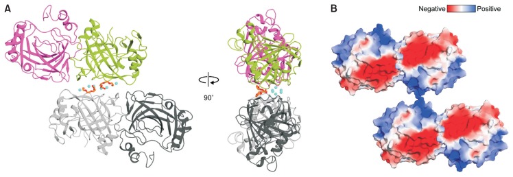
Fig. 3. Crystallographic interface of the pmCA.
(A) Two possible dimers in crystal of pmCA. Crystal packing of pmCA in the C2 space group (crystal form 1) is almost identical with those of pmCA in the P21212 space group (crystal forms 2 and 3). Two monomers and their two-fold symmetry-related molecules are colored magenta, green, dark and light gray, respectively. Calcium ions and PEG molecules bound at the crystallographic interface are shown as cyan spheres and orange sticks, respectively. (B) Electrostatic surface potential of pmCA. The crystallographic interface is split and rotated by 90°. White, neutral; blue, positively charged; red, negatively charged. Surface electrostatic potential was calculated using the PDB2PQR and APBS server (http://www.poissonboltzmann.org).

