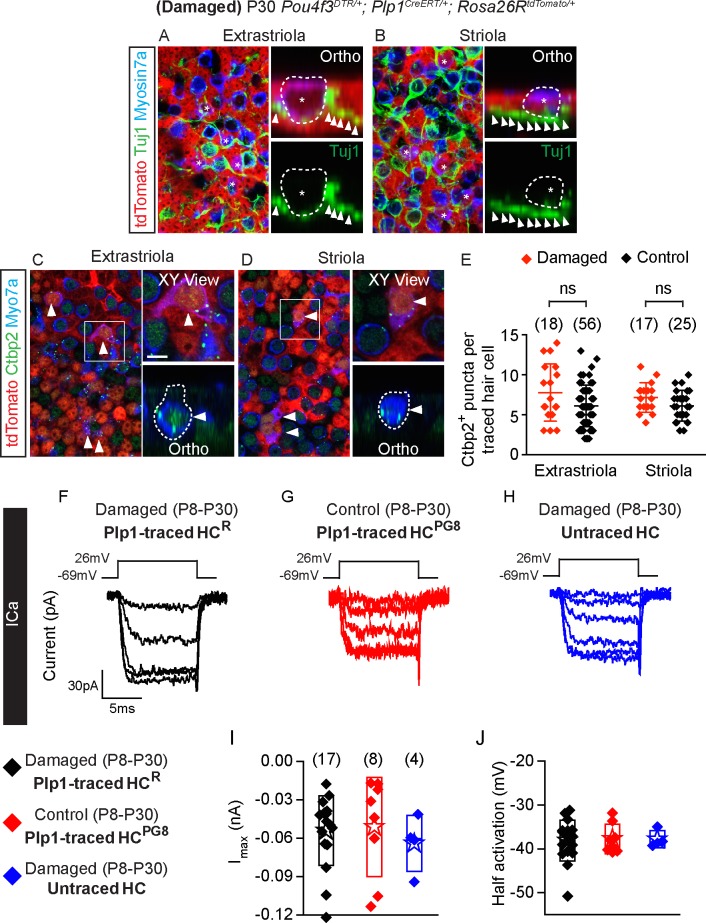
Fig 8. Synaptic properties of Plp1-traced, regenerated hair cells.
A-B) Representative images of regenerated tdTomato+/Myosin7a+ hair cells (asterisks) with associated with Tuj1+ neural elements (arrowheads) in the extrastriola and striola (n = 106 cells from 5 mice). C-D) All Plp1-traced hair cells examined expressed Ctbp2 on the basolateral surfaces (arrowheads, n = 35 cells from 3 mice). E) Quantification of Ctbp2+ puncta in traced hair cells in the extrastriola and striola. No significant difference was found between hair cells from control and damaged utricles (n = 56 extrastriolar and 25 striolar hair cells from 6 control mice utricles, n = 18 extrastriolar and 17 striolar hair cells from 3 damaged mice utricles). F-H) Representative calcium currents from traced hair cells (HCR from damaged utricles and HCPG8 from undamaged utricles) and untraced hair cells. Calcium currents were isolated using methods described in Fig 3. I-J) Peak current responses and maximal current and half activating voltage were not statistically different among groups (n = 4–17 cells). Data shown as mean ± SD, compared using Student t tests and one-way ANOVA by Kruskal Wallis-Dunn's multiple comparison tests. Scale bars: A-D) 20 μm, XY view) 5 μm. The underlying data can be found within S1 Data. HCPG, postnatally generated hair cell.

