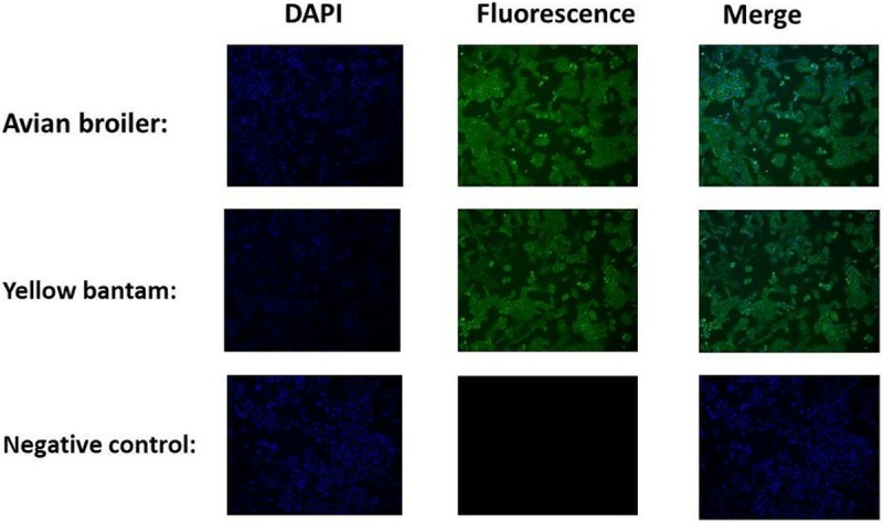
Fig 1. Immunofluorescence of markers in cartilage cells.

Nuclei stained with DAPI are shown in the left panels. The pictures above indicated that staining of the cells for the marker collagen II was positive. The merged images are shown in the right-most panels. Scale bar = 100 μm.
