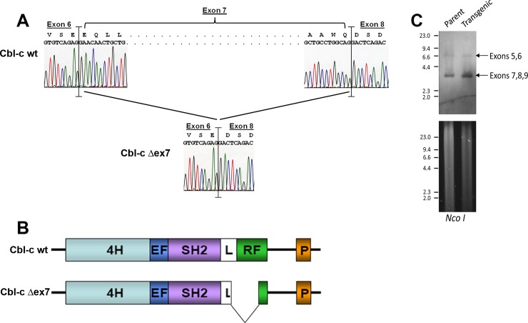
Fig 1. Structure of a Cbl-c mutant (Cbl-c Δex7) isolated from a mouse ductal carcinoma.
(A) Sequence analysis of Cbl-c wildtype and Cbl-c Δex7. Sequence for wild type cDNA is shown on top of panel and sequence for mutant is shown on bottom demonstrating an in-frame deletion of exon 7 which encodes part of the linker domain and the catalytic RING finger domain. (B) Cartoon depicting structural comparison of mouse wild-type and mutant mouse Cbl-c Δex7. (C) Genomic DNA from parental and transgenic mouse stocks were digested, as indicated, separated by electrophoresis, stained with cyber green (bottom panel), then transferred to nylon membrane (upper panel) and probed to detect regions flanking exon 7 of mouse Cbl-c. Arrows indicate the bands containing exons 5 and 6 and the band containing exons 7-9.DNA molecular weight standards in kilobases are indicated on the left of each panel.

