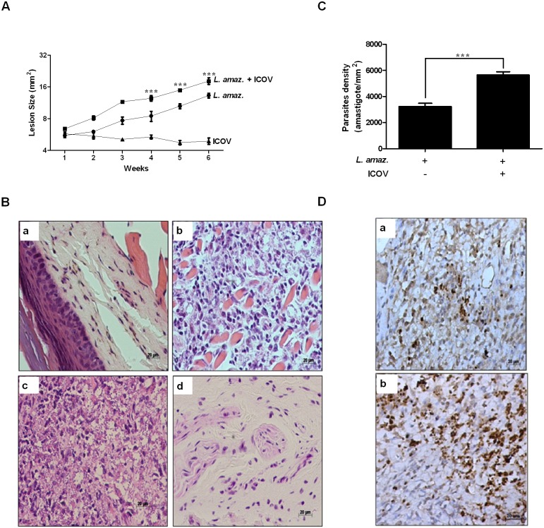
Fig 4. Coinfection of mice with Icoaraci exacerbates the infection of Leishmania (L.) amazonensis.
(A) Wild-type C57BL/6 mice were infected with stationary-phase promastigotes of L. (L.) amazonensis at a ratio of 5 x 10⁶, 2,25 x 10⁵ PFU of Icoaraci virus, or coinfected. The diameter of the paw was evaluated once a week and expressed as the difference between the size of the infected and uninfected paws. (B) In the fifth week after infection, a histopathological analysis of skin lesions of mouse footpads was performed (a—without infection, b—L. (L.) amazonensis infection, c—L. (L.) amazonensis + ICOV infection and d—ICOV infection). (C) Graphical analysis of reactive cells positive for Leishmania. (D) The density of amastigotes was evaluated by immunohistochemical reaction with antibody against Leishmania (a—L. (L.) amazonensis infection, b—L. (L.) amazonensis + ICOV infection). Images are representative of 5 animals in different groups. Asterisks indicate significant differences between groups by ANOVA or Student’s t-test, with *** p <0.0001.

