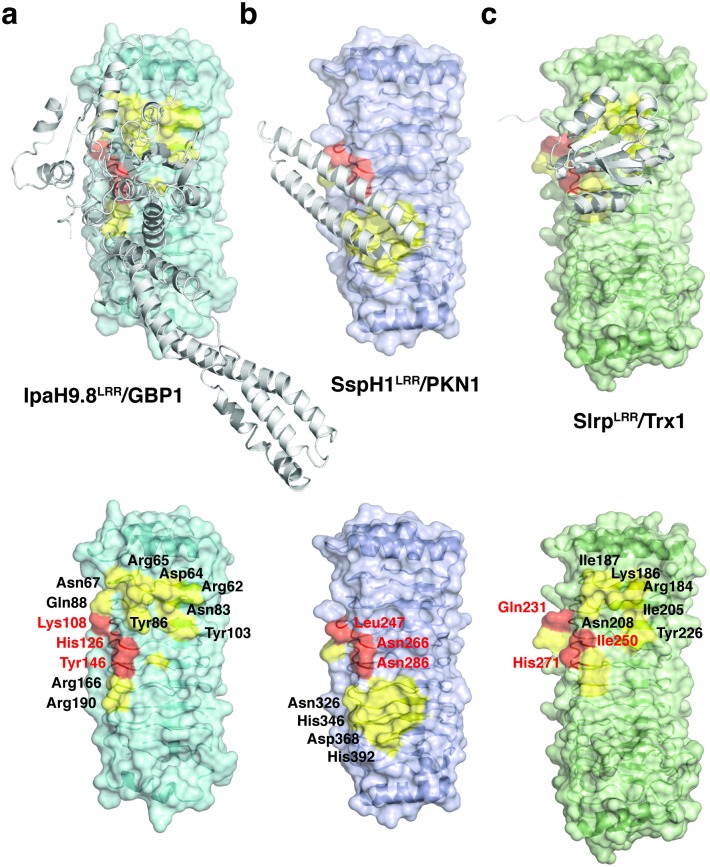
Fig 5. Interaction hot spots in the LRR domains of the IpaH family proteins.
(a) The structure of IpaH9.8LRR in complex with GBP1. IpaH9.8LRR is shown as colored ribbon diagrams enclosed in its van der Waals surface. GBP1 is shown as white ribbons. IpaH9.8 residues involved in binding to GBP1 are colored in yellow, and the three hot spot residues are highlighted in red. A IpaH9.8LRR alone structure without GBP1 is shown at the bottom for the GBP1-binding residues to be clearly seen. (b) The structure of SspH1LRR in complex with a coiled-coil region of the PKN1 kinase (PDB ID: 4NKG). SspH1LRR is shown as colored ribbon diagrams in its van der Waals surface, and PKN1 is shown as white ribbons. (c) Type I binding site in the structure of SlrpLRR in complex with human Trx1 thioredoxin (PDB ID: 4PUF). SlrpLRR is shown as colored ribbon diagrams in its van der Waals surface, and Trx1 is shown as white ribbons.

