Fig. 1.
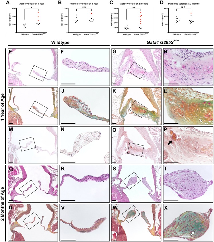
Gata4G295Ski/wt mice exhibit semilunar valve stenosis. (A-D) Valve stenosis is found at 1 year (A,B) and 2 months of age (C,D) in Gata4G295Ski/wt mice compared with wild-type controls as determined by echocardiography (1 year old mice: n=7 for wild type and n=5 for Gata4G295Ski/wt; 2 month old mice: n=9 for wild type and Gata4G295Ski/wt). Velocity from Gata4G295Ski/wt mice indicated as squares, wild-type mice are shown as circles. Red circles or squares indicate stenosis. (E-L) Histological sections of aortic valve of Gata4G295Ski/wt mice exhibit thickened, dysmorphic leaflets (G,H) and abnormal ECM, as shown by Russell-Movat's Pentachrome staining (K,L) at 1 year of age, compared with wild-type control valves (E,F,I,J). F,H,J and L are high magnification images of the boxed areas in E,G,I and K, respectively. (M-P) Positive Alizarin red staining consistent with a calcific nodule (black arrow) is noted at 1 year of age (O,P) compared with wild-type control (M,N). P and N are high magnification images of the boxed areas in O and M, respectively. (Q-X) Aortic valve sections from 2 month old Gata4G295Ski/wt mice demonstrate thickened, dysmorphic leaflets (S,T) and abnormal ECM (W,X) compared with wild-type control valves (Q,R,U,V). White arrow highlights proteoglycan-rich nodule formation. R,T,V, and X are high magnification images of the boxed areas in Q,S,U and W, respectively. For each histological section shown in E-X, n=3 for wild type and n=3 for Gata4G295Ski/wt. For Russell-Movat's Pentachrome stains, yellow indicates elastin and blue indicates proteoglycans. *P≤0.05; **P≤0.005; N.S., P >0.05 (Student's t-test). Scale bars: 100 µm.

