Fig. 1.
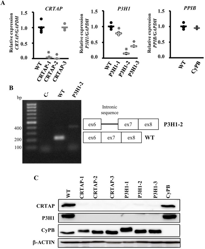
Loss of mutant CRTAP, P3H1 and CyPB in OI patient fibroblasts. (A) Quantitation of CRTAP, P3H1 and PPIB expression evaluated by qPCR. Mutations in CRTAP, P3H1 and PPIB caused a close to complete absence of the mutated transcripts in CRTAP-1, CRTAP-2 and P3H1-2 patients, and a reduced mRNA level in P3H1-1 and P3H1-3. *P<0.05. WT values are represented as black dots; CRTAP as gray dots; P3H1 as white dots; CyPB as dark gray dots. (B) Amplification of the exon 6-exon 8 region of P3H1 transcript generated the expected 217 bp amplicon in control cells (WT), whereas, in the P3H1-2 patient, the presence of a higher molecular weight (∼400 bp) band compatible with intronic retention was detected. C-, RT-PCR negative control. (C) Representative western blot to evaluate the expression of CRTAP, P3H1 and CyPB in control (WT) and mutant cell lysate fractions (CRTAP-1, CRTAP-2, CRTAP-3, P3H1-1, P3H1-2, P3H1-3, CyPB). Loss of the mutated protein in patient's cells was demonstrated. Patients with mutations in CRTAP showed also no P3H1 expression and patients with mutations in P3H1 showed no CRTAP expression, as a consequence of their mutual protection in the complex.

