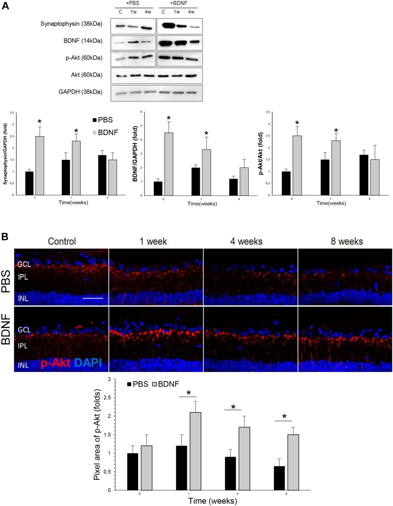
Fig. 2.
Effects of BDNF application to expression of synaptic proteins and phosphorylated Akt. (A) Results of western blot analysis of BDNF, synaptophysin and phosphorylated Akt. There were significant elevations in BDNF in the BDNF-injected group compared with the PBS-injected group at baseline and 1 week after cauterization. Additionally, there was a significant increase in the level of synaptophysin, which is a synaptic vesicle protein, and phosphorylated Akt in the retinas of the BDNF-injected group compared with the PBS-injected group at baseline and 1 week after BDNF-injection. For the western blot analysis, the PBS-injected group and the BNDF-injected group each included 6 retinas; total n=36. *P<0.05. (B) Confocal micrographs of retinal sections stained for phosphorylated Akt. In the BDNF-injected group, there was an increase in phosphorylated Akt levels in the innermost IPL compared with the PBS-injected group; this peaked at week 1 and lasted until week 8. For the p-Akt staining and quantification, the PBS-injected group and the BDNF-injected group each included 6 retinas at each time point; 10 sections per retina were analyzed; total n=48. Scale bar: 50 μm. *P<0.05.

