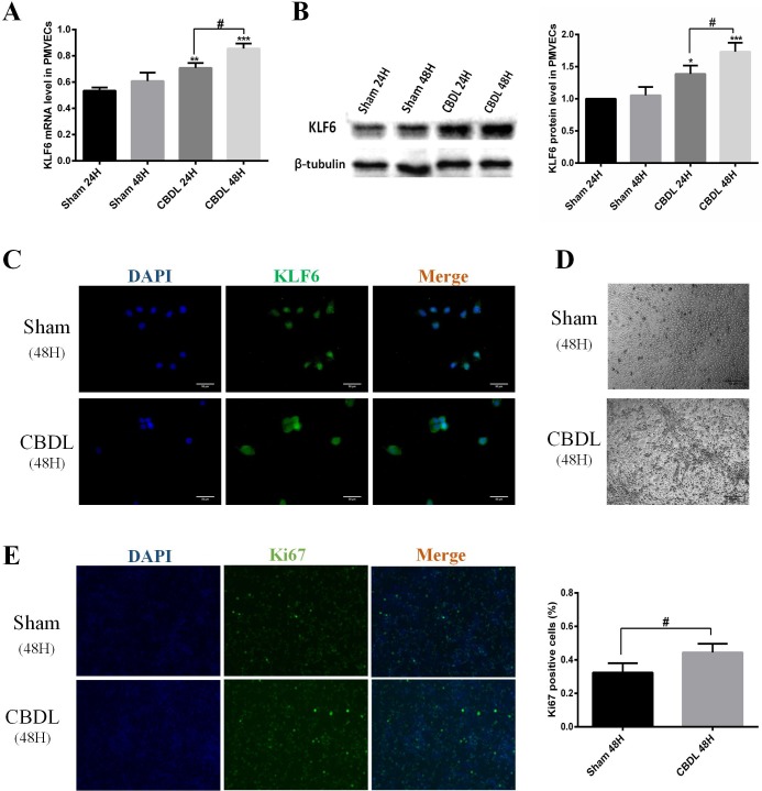
Fig. 3.
KLF6 expression was induced in vitro of the HPS model. (A) Representative mRNA levels of KLF6 in PMVECs with sham or CBDL serum stimulated for 24 h and 48 h (n=3). (B) Representative immunoblots and graphical summaries of KLF6 levels in PMVEC-exposed sham or CBDL serum in 24 h and 48 h (n=3). (C) Representative immunofluorescence images of KLF6 in PMVECs under the condition of sham serum or CBDL serum for 48 h (n=3). (D) Representative micrographs of PMVECs exposed to sham or CBDL serum for 48 h. (E) Representative immunofluorescence staining of ki67 by positive proliferation PMVECs under the condition of sham serum or CBDL serum for 48 h (n=3). Data are presented as the mean±s.d., *P<0.05, **P<0.01 ***P<0.001, compared with the sham 24-h group; #P<0.05 compared with the labeled groups.

