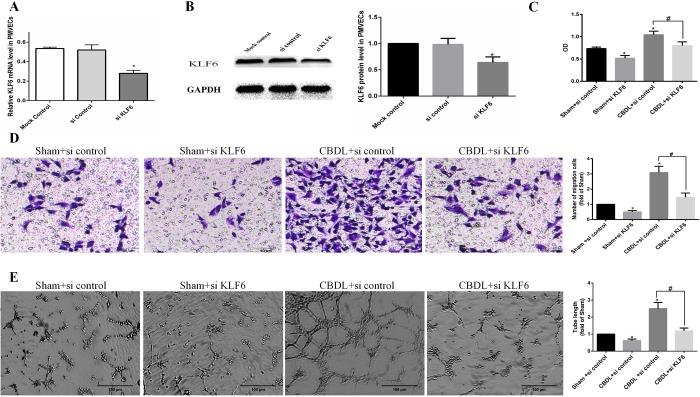
Fig. 4.
Involvement of KLF6 in PMVECs mediated the proliferation, migration and tube formation in an in vitro HPS angiogenesis model (n=3). (A,B) PMVECs pretreated with control siRNA (siControl, 100 nM) or siKLF6 (siKLF6, 100 nM). SiKLF6 efficiently reduced KLF6 mRNA (A) and protein (B) levels. (C) Pretreated PMVECs were seeded in 96-well plates with the same medium and cultured for 24 h. Cell proliferation was determined by the Cell Counting Kit-8 assay and by measuring the absorbance at 450 nm. OD, optical density. (D) Pretreated PMVECs were seeded and cultured in an upper chamber with the same media in a lower chamber for 24 h, and the number of PMVECs that migrated to the lower chamber was counted. (E) Pretreated PMVECs were seeded and cultured in Matrigel, and tube length was measured after 8 h. Values are expressed as the mean±s.d. *P<0.05 compared with the sham+si control group; #P<0.05 compared with the CBDL+si control group.

