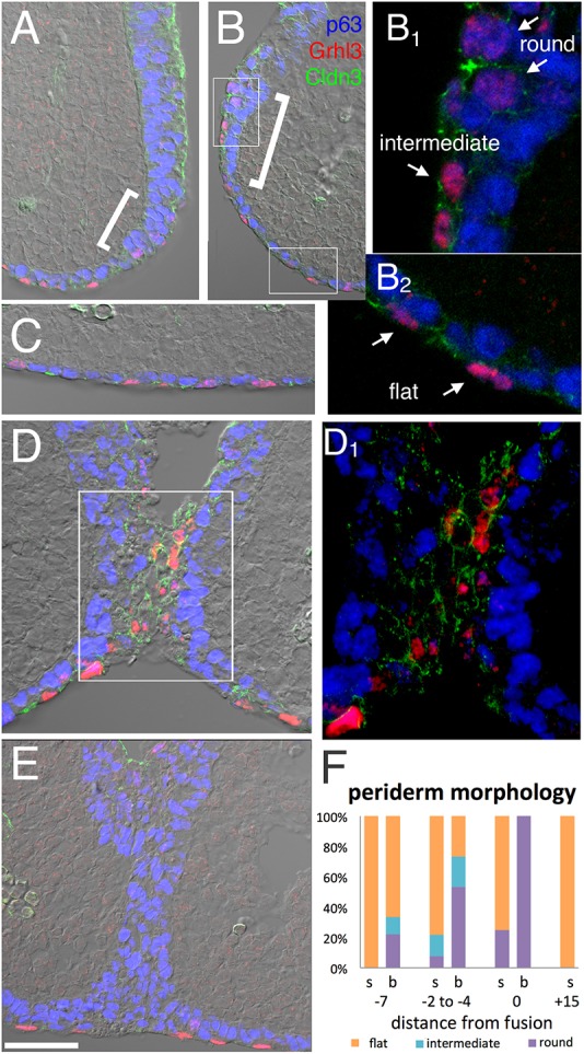
Fig. 8.

Periderm around the fusing lambdoid junction. (A-E) Immunofluorescent imaging of Grhl3 (red) in periderm nuclei, p63 (blue) in basal cell nuclei and Cldn3 (green) in cellular junctions of periderm, OE and endothelial, but not basal, cells overlaid onto a DIC image to visualize tissue organization. (A,B) The prospective fusion zone of the MNP and LNP two sections anterior (distal) to the contact point (shown in D). The brackets indicate the transition zone (the prospective fusion zone) between surface ectoderm and nasal pit ectoderm. White boxes in B indicate the surface epithelium and border epithelium, and are shown at higher magnification in B1 and B2, respectively, with arrows indicating examples of round, flat and intermediate Grhl3+ nuclei. (C) Surface epithelia near the facial midline, where p63+ basal cells and Grhl3/Cldn3+ periderm form an ordered bilayer. (D) Contact point where Cldn3/Grhl3+ cells bridge the gap, with the boxed area shown at higher magnification in D1. (E) The epithelial seam 15 sections posterior to the fusion point. Seam has p63+ cells but lacks Grhl3+ cells. (F) Quantification of periderm nuclear morphologies in surface epithelia (s) or prospective fusion zone (b), with distances from the fusion site indicated by relative section number. Scale bar: 50 µm in A,B,C,D,E.
