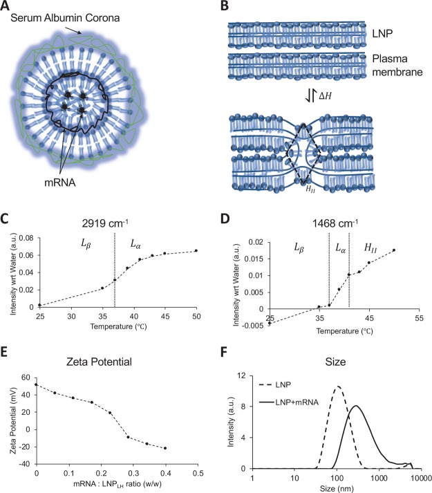
Figure 1.
Physical characteristics of the lipid nanoparticle. (A) Schematic of the LNP–mRNA complex. Amphiphilic lipid molecules are drawn to show the lamellar fluid phase (Lα) and inverted hexagonal (HII) phase coexistence. mRNA molecules assemble in the intralamellar space of the Lα phase or the hydrophilic core of the HII phase. When incubated with plasma, a serum albumin corona forms around the shells shielding the positive charge of the nanoparticle. (B) Intermediate stalk formation during the fusion of two lamellar bilayers is shown take place via the (HII) phase. The transition occurs via reversible exchange of enthalpy ΔH. (C) Peak intensity in FTIR spectra of the LNP at 2919 cm–1 measured as a function of temperature. The peak corresponds to the stretching of −CH2 bonds and identifies the gel (Lβ) to fluid (Lα) transition near 37 °C.27 (D) Peak intensity in FTIR spectra of the LNP at 1468 cm–1 measured as a function of temperature. The peak corresponds to the scissoring of −CH2 bonds and identifies two kinks corresponding to gel (Lβ) to fluid (Lα) transition near 37 °C and lamellar fluid (Lα) and inverted hexagonal (HII) transition near 41 °C.27 (E) Zeta potential of LNP/mRNA lipoplex as a function of mRNA titrated reported as the w/w ratio on x axis. The w/w ratio of 0.2 in the vicinity of the isoelectric point was used as initial estimate for the optimum mRNA to LNP ratio (F) size distribution of the lipid nanoparticles measured using a Zetasizer before and after association with the native mRNA at the w/w ratio of 0.2 (zeta potential 19.1 mV).

