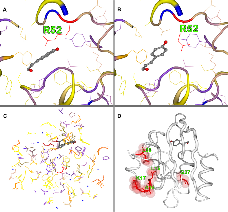
Figure 2.
2StrucCompare screenshot downloads for the structure intermediate IL1 (1T1A) from the photoactive yellow protein (PYP) compared with the dark structure (1T18.B). (A) The dark conformation of Arg52 adjacent to the chromophore site. (B) The IL1 Arg52 conformation where the side chain has been ejected from adjacent to the chromophore. (C) A paired view of the side chain only, showing the large numbers of residues that have an increased difference in their positions in the IL1 intermediate from that of their initial dark positions. (D) The differences (‘diff’) representation of 2StrucCompare showing the positions of difference between the assigned secondary structures of the dark and IL1 states. This indicates that small conformational changes are already arising in residues near the N-terminal of the IL1 intermediate as a result of the photoexcitation.

