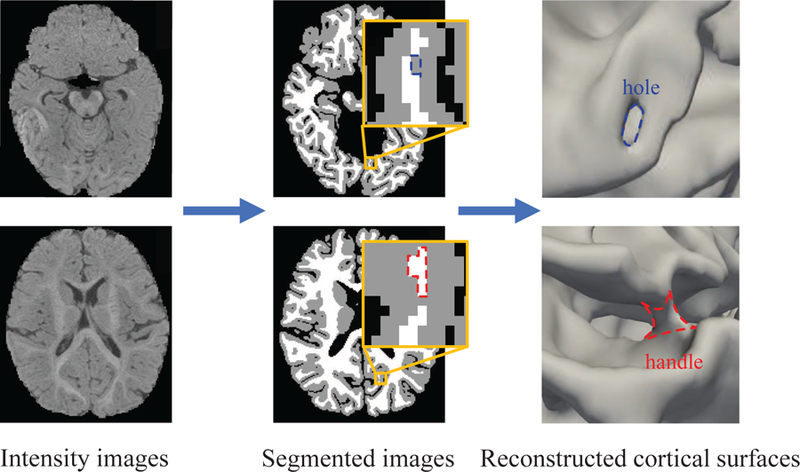
Figure 1.

Illustration of topological errors in the tissue segmentation result of a 6-month-old T1-weighted MRI. Although the image was segmented automatically by an infant-dedicated tissue segmentation method Wang et al. (2018), it still inevitably contains many segmentation errors, which lead to abounding topological errors on the reconstructed cortical surfaces. The blue and red contours in the segmented image and cortical surface indicate topological defects (with the blue contour indicating a hole and the red contour indicating a handle).
