
Figure 5.
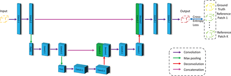
The architecture of our proposed Anatomically Constrained U-Net (AC-U-Net) method. The purple, green, red and peach arrows represent the convolution, max pooling, deconvolution, and concatenation operations, respectively. The orange, red, yellow and green patches represent the input, output, ground truth and atlas reference, respectively.