
Figure 9.
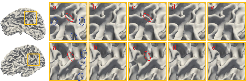
Visual inspection of the original white matter surfaces (a), manually corrected surfaces (b), and AC-U-Net corrected surfaces at the 1st iteration (c), 2nd iteration (d), and 3rd iteration (e) on human infant MRI dataset with simulated topological errors, respectively. The red and blue circles represent handles and holes, respectively.