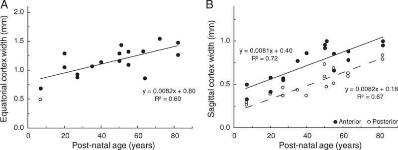
Figure 4.

Human lens cortical thickness as a function of age. Thickness was determined from the distance between the outer boundary of the 30 mg nucleus and the lens capsule for (A) the equatorial cortex and (B) the anterior (●) and the posterior (○) sagittal cortex. The open symbol in Figure 4A is an outlier, which differed from the mean for that age by more than 2.5 times the standard deviation. It was not used in the regression analysis.
