
Figure 4.
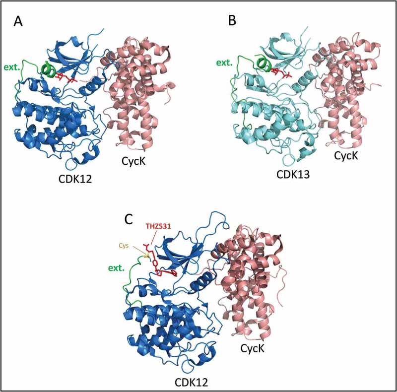
Crystal structures of recombinant human CDK12/13•Cyclin K complexes.
(a) Green color labels the segment of CDK12 chain that extends out from the back (C-term) of the well-folded kinase domain, as discussed in the text. The last residue of the short helix at the end of the green segment is Lys1046. The ADP in the active site is colored red. From PDB ID 4NST; image using PyMol. (b) Green color labels the segment of CDK13 chain that extends out from the back (C-term) of the well-folded kinase domain, as discussed in the text. The ADP in the active site is colored red. From PDB ID 5EFQ; image using PyMol. (c) Inhibitor THZ531 (red) in active site of CDK12 & extending out to react with Cys1039 (yellow). PDB ID 5ACB; image using PyMol.