
Figure 1.
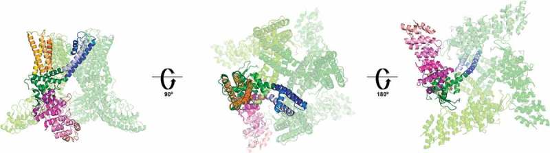
Structure of the TRPV1 channel. Only one of the subunits is highlighted for clarity.
The ankyrin repeat domain is shown in purple, the S1- S4 domain in orange, the pore forming S5-P-S6 is shown in blue. The pre-S1, S4-S5 linker, and TRP domain, which participate in the allosteric modulation of the channel, are shown in green. The left panel is a lateral view, the central and right panels are extracellular and intracellular views, respectively. (PDB 3J5P).