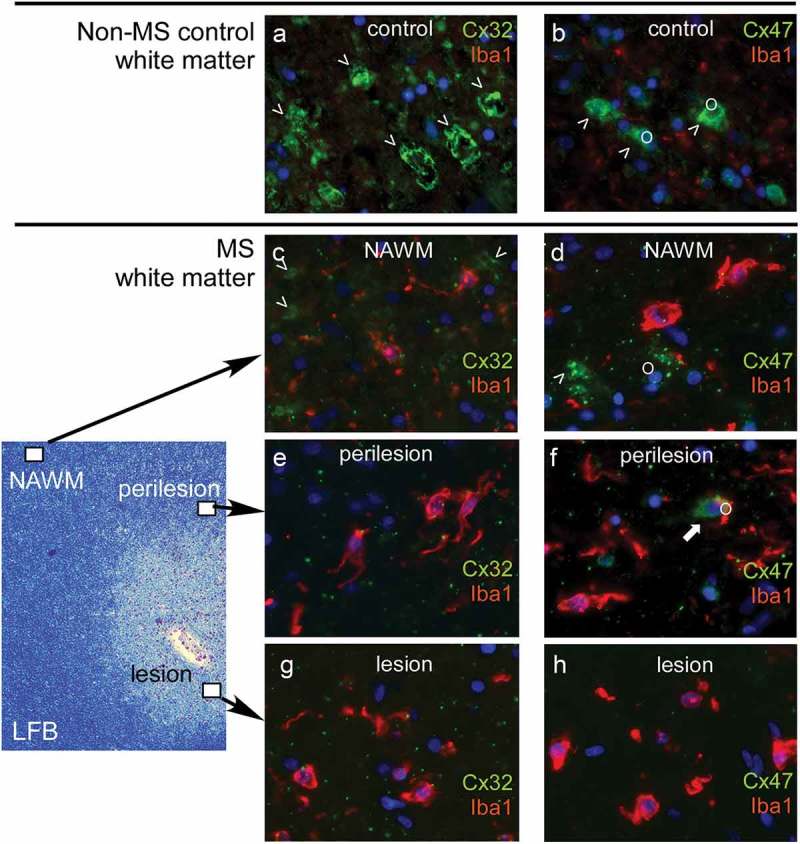
Figure 2.

Expression of oligodendrocyte connexins in multiple sclerosis (MS) brain. Images of the white matter of a non-multiple sclerosis control (a- b) compared to a multiple sclerosis patient (c- h) are shown. Low magnification of MS brain white matter stained with Luxol fast blue (LFB) is shown on the left to indicate the location of the high magnification immunofluorescence pictures shown on the right. Antibodies to oligodendrocyte connexins as indicated (green) were combined with the Iba1 antibody labeling microglia (red). Cell nuclei are stained blue. The normal expression of Cx32 along large myelinated fibers in non-MS control brain (a) is significantly reduced, not only within and around lesions but also in NAWM in MS brain (c, e, g), associated with prominent microglia activation. Likewise, Cx47 expression mainly in cell bodies and proximal processes of oligodendrocytes (o) shown in non-MS brain (b) is reduced in and around MS lesions whereas, in contrast to Cx32, it appears preserved in NAWM (d, f, h).
