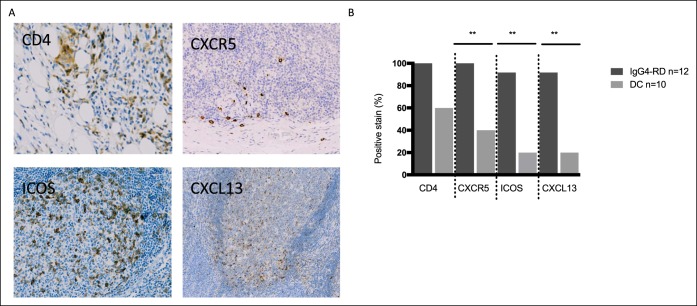
Figure 6.
CD4+ T cells and Tfh cells are present in IgG4-RD tissue specimens and home to lymphoid follicles. Paraffin-embedded tissue specimens were stained for Tfh-cell markers. (a) Representative panels from patients with IgG4-SC/AIP stained for CD4 (liver, top left panel), CXCR5 (pancreas, top right panel), ICOS (pancreas, bottom left panel), and CXCL13 (pancreas, bottom right panel). Positively staining cells stain DAP-positive (brown). (b) Histogram shows the percentage of positively staining specimens for CD4+, CXCR5+, CXCR5+ICOS+, and CXCL13+ cells. Data from 12 patients with IgG4-SC/AIP (red bars) and 10 disease controls (gray bars). **P values as in Figure 2. AIP, autoimmune pancreatitis; DC, disease control; IgG4-RD, IgG4-related disease; IgG4-RI, IgG4–responder index; IgG4-SC, IgG4-related sclerosing cholangitis.

