Figure 5.

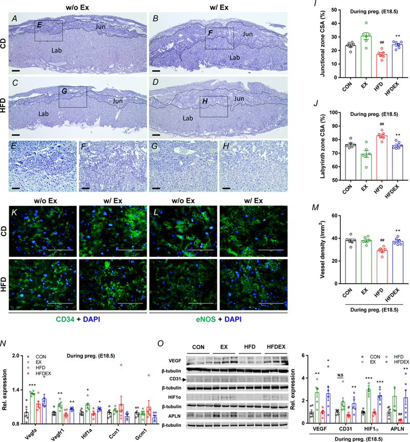
Maternal exercise training reverses impaired placental vascularization in HFD-mice. A-H,K,L, Representative images of hematoxylin and eosin (H&E), CD34 and eNOS immunocytochemistry (ICC) staining of placenta from CON or HFD mice with/without exercise, Scale bar, 500 μm (A-D), 100 μm (E-H,K,L), junctional zone (Jun) and labyrinth zone (Lab). I-J, Cross-sectional area (CSA) of junctional zone (I) and labyrinth zone (J) at E18.5 in placenta. M, Vessel density per mm2 in labyrinth zone of placenta. N, mRNA levels of different vasculogenic markers in CON or HFD mice with/without exercise. Expression was normalized by ΔCt values. O, cropped western blots of VEGF, CD31, and APLN in the placenta (β-tubulin was used as the loading control) from CON or HFD with/without exercise. Data are expressed as the mean ± s.e.m. n = 6. *P < 0.05, **P < 0.01, and ***P < 0.001 in CON vs. EX or HFD vs. HFDEX; ##P <0.01 in CON vs. HFD by two-tailed Student’s t-test followed by one-way ANOVA with post hoc Bonferroni multiple comparison analysis (I-O).
