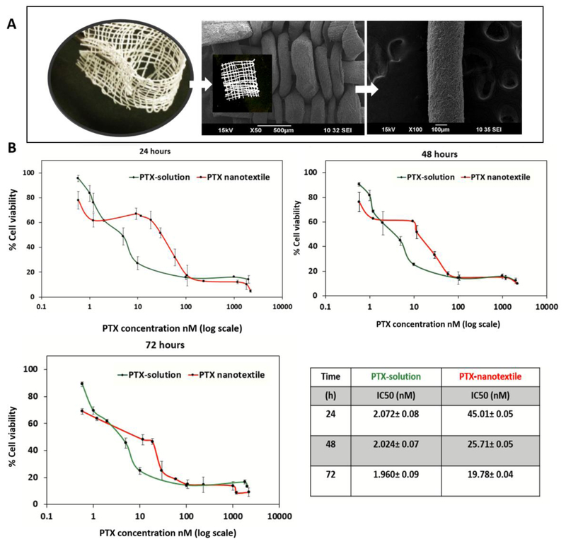
Figure 1. Nanotextile implant and in vitro cytotoxicity of PTX in ID8-VEGF cells.
(A) Optical and scanning electron microscopy images of paclitaxel (PTX)-loaded polydioxanone (PDS) yarn and woven nanotextile implant (inset image) (B) The plots of ID8-VEGF cell viability versus PTX concentration administered in solution and from nanotextiles incubated for 24, 48 and72 h. The PTX IC50 values were calculated and shown in the table.

