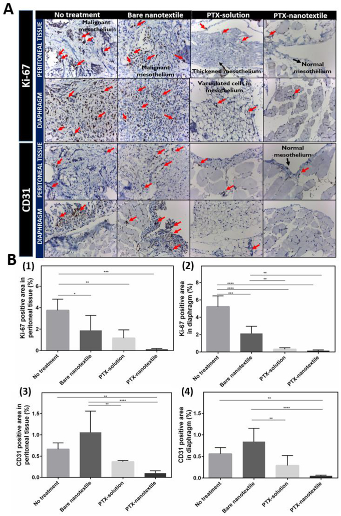
Figure 4. Expression of proliferation and angiogenic markers.
(A) Immunohistochemistry (IHC) microscopic analysis at 40X magnification showing the expression of proliferation (Ki-67) and angiogenic (CD31) markers in formalin- fixed, paraffin-embedded peritoneal tissue and diaphragm of mice from control and treatment groups euthanized on day 35. (B) Semiquantitative analysis of diaminobenzidine stained positive area from the IHC images for Ki-67 and CD31 expression using NIH Image J software.

