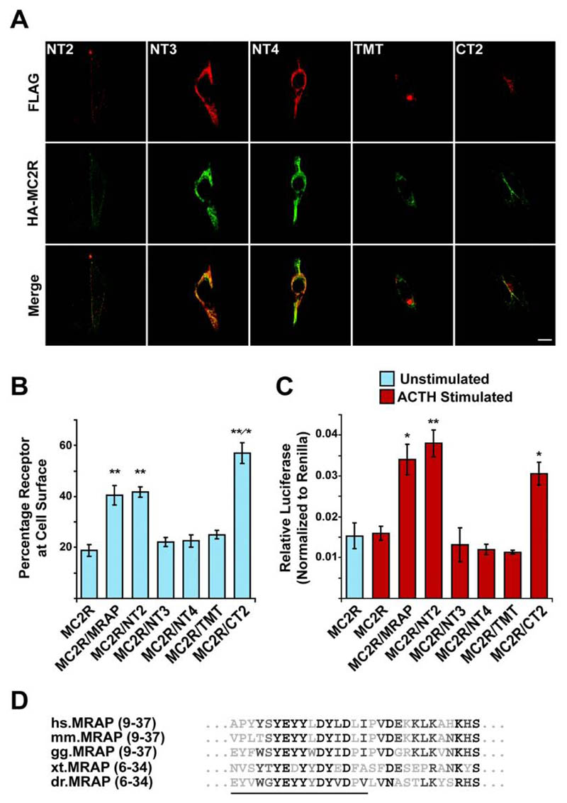
Figure. 4. MC2R cell surface trafficking requires the N-terminus of MRAP and is regulated by the C-terminus of MRAP.
(A) Immunofluorescent staining showing localization of HA-MC2R with each of the FLAG tagged MRAP constructs in permeabilized transiently transfected CHO cells. Top panels, anti-FLAG staining of MRAP constructs (red). Middle panels, anti-HA staining of HA-MC2R (green). Bottom panels, merge. HA-MC2R co-localizes with each of the MRAP constructs, and is detected at the cell surface in the presence of the NT2 and CT2 constructs. With NT3, NT4, and TMT, HA-MC2R is not seen at the cell surface. Bar, 10μm. (B) Fluorescent cell surface assay showing the percentage of MC2R at the cell surface when co-transfected with each of the MRAP constructs. Significant cell surface expression is seen in the presence of MRAP, NT2, and CT2 (**, P < 0.005) compared to HA-MC2R alone. There is no increase seen with the remaining constructs. HA-MC2R cell surface expression is significantly higher when co-expressed with CT2 compared to MRAP or NT2 (*, P < 0.05). Mean ± SEM; n=4. (C) Luciferase assay to measure cAMP response following 6 hour 10-6 M ACTH stimulation. There is a significant increase in cAMP following ACTH stimulation when HA-MC2R is co-expressed with MRAP, NT2, and CT2. Mean ± SEM; n=3; * P < 0.05, **, P < 0.005. (D) Alignment of amino acids 9 to 37. Residues deleted in the NT3 construct are underlined. hs, Homo sapiens. mm, Mus musculus. gg, Gallus gallus. xt, Xenopus tropicalis. dr, Danio rerio.

