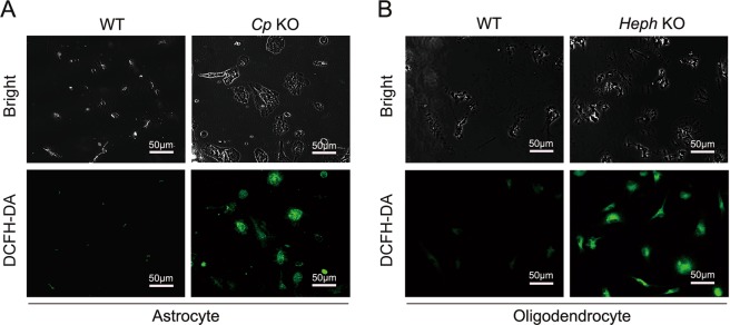
Figure 3.
Intracellular oxidative stress in Cp KO astrocytes and Heph KO oligodendrocytes after iron treatment. WT and Cp KO astrocytes (A), and WT and Heph KO oligodendrocytes (B) were loaded with iron for 24 hours, then incubated for a further 24 hours in medium without iron. The cells were then stained with DCFH-DA to detect reactive oxygen species. Representative brightfield (top) and fluorescent (bottom) images are shown. Data are representative of at least three independent experiments.

