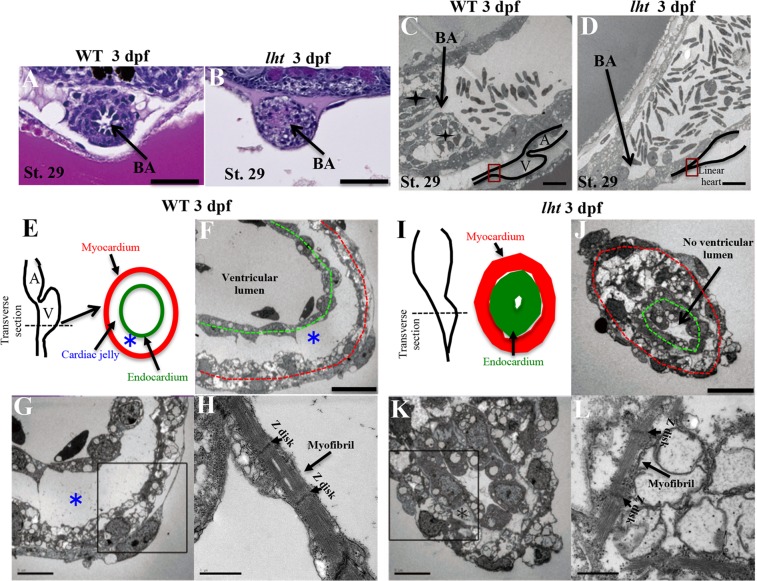
Figure 3.
The lht mutant lacks cardiac jelly and displays a constricted outflow tract. Haematoxylin and eosin (H&E) staining of WT and lht mutant hearts at 3 dpf in the outflow tract displaying differences in cellular arrangement and lumen development (n = 3) (A,B). Scanning electron micrographs of a sagittal section of the BA, ✦ represents the cellular arrangement around the outflow tract in WT (n = 2) (C,D). Schematic representations of a transverse section of the heart from WT and lht mutant embryos (E,I). Black dotted line indicates parts of the sections represented in (F–H and J–L). Scanning electron micrograph of the transverse section of the ventricular chamber of a WT embryo and the distal part of the linear heart tube of an lht mutant embryo at 3 dpf (n = 2) (F,G and J,K). The myocardium and endocardium are represented by red and green dotted lines, respectively; *indicates cardiac jelly. Myofibril ultrastructure of the heart of WT (H) and lht mutant embryos (L). BA, bulbus arteriosus. Scale bars: 50 μm (A,B), 10 μm (C,D,F,J), 5 μm (G,K), and 1 μm (H,L).

