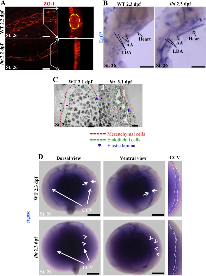
Figure 6.
The lht mutant embryo exhibits defects in vascular maturation. Whole-mount immunohistochemistry for ZO-1 (a tight junction protein) at 2.2 dpf for WT and lht mutant embryos (A). Images are 3D projections of z-stack images acquired by confocal microscopy. Inset images display lumen formation in a blood vessel. Yellow circle depicts the vascular lumen in WT embryos (n > 10). In situ hybridization for EGFL7 expression in WT and lht mutant embryos at 2.3 dpf (n > 10) (B). Transmission electron micrograph of a transverse section of the dorsal aorta of WT and lht mutant embryos at 3.1 dpf (C; n = 2). In situ hybridization for etgase at 2.3 dpf (D). White arrows pointing at the CCV in WT represent the wide vascular lumen surrounded by etgase-positive cells. The white arrowheads in lht mutant embryos show disoriented etgase-positive cells around a putative blood vessel (n > 10). White dotted lines in the right-most panel indicate the vascular lumen. AA, aortic arch; LDA, lateral dorsal aorta; etgase, embryonic transglutaminase. Scale bars: 20 μm (A), 100 μm (B), 250 μm (C), 10 μm (B).

