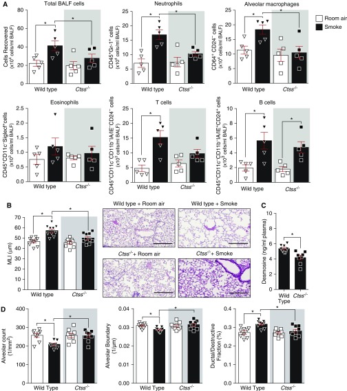
Figure 3.
Ctss (cathepsin S) deficiency prevents smoke-induced lung immune cell infiltration and airspace enlargements in mice. Wild-type and Ctss−/− mice were exposed to room air and cigarette smoke for 6 months. (A) BALF total immune cells, neutrophils, alveolar macrophages, eosinophils, T cells, and B cells were quantified in each group by flow cytometry. (B) Mean linear intercepts were measured in the lungs of the mice to assess air space size, and comparative histology images of the four mouse groups are presented here (scale bars, 40 μm). (C) Plasma desmosine levels were determined in smoke-exposed animals by ELISA. (D) Alveolar count, alveolar boundary, and ductal/destructive fractions were quantified in each animal by parenchymal airspace profiling. Data are represented as mean ± SEM, where n ≥ 5 per group. *P < 0.05, when comparing both treatments connected by a line, determined by two-way ANOVA with Tukey post hoc test or Student’s t test when comparing only two groups. BALF = BAL fluid; MLI = mean linear intercept.

