Figure 7.
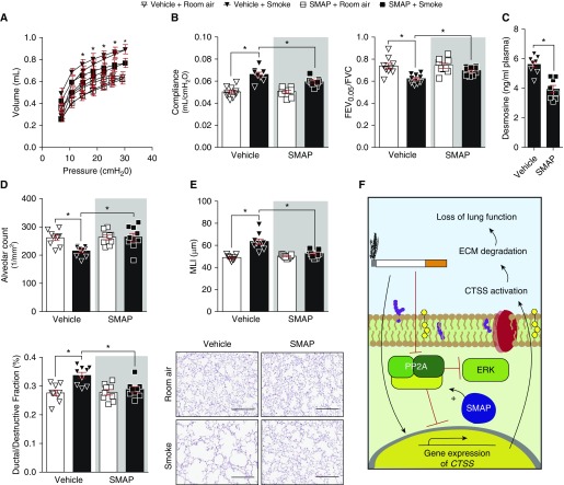
Activating PP2A (protein phosphatase 2A) signaling prevents smoke-induced loss of lung function. Mice were exposed to room air and cigarette smoke and two oral administrations of small molecule activator of PP2A daily for 2 months. (A and B) Negative pressure-driven forced expiratory and forced oscillation technique maneuvers were performed in all animal groups. (A) Pressure–volume loops and (B) compliance and forced expiratory volume in 0.05 seconds (FEV0.05)/FVC were determined in each animal. (C) Plasma desmosine levels were assessed in smoke-exposed animals by ELISA. (D) Alveolar count and ductal/destructive fractions were quantified in each animal by parenchymal airspace profiling. (E) Mean linear intercepts were measured in the lungs of the mice to assess air space size, and comparative histology images of the four mouse groups are presented here (scale bars, 40 μm). Data are represented as mean ± SEM, where n = 9 per group. *P < 0.05, when comparing both treatments connected by a line, determined by two-way ANOVA with Tukey post hoc test. (F) Illustration of the possible signaling mechanism for PP2A regulation of CTSS (cathepsin S). Evidence presented in this study indicates that PP2A prevents signaling leading to CTSS gene expression, but after smoke exposure, CTSS expression is enhanced and the phosphatase activity of PP2A is diminished. This enhancement of CTSS directly impacts lung function. ECM = extracellular matrix; ERK = extracellular signal–regulated kinase; MLI = mean linear intercept; SMAP = small molecule activator of PP2A.

