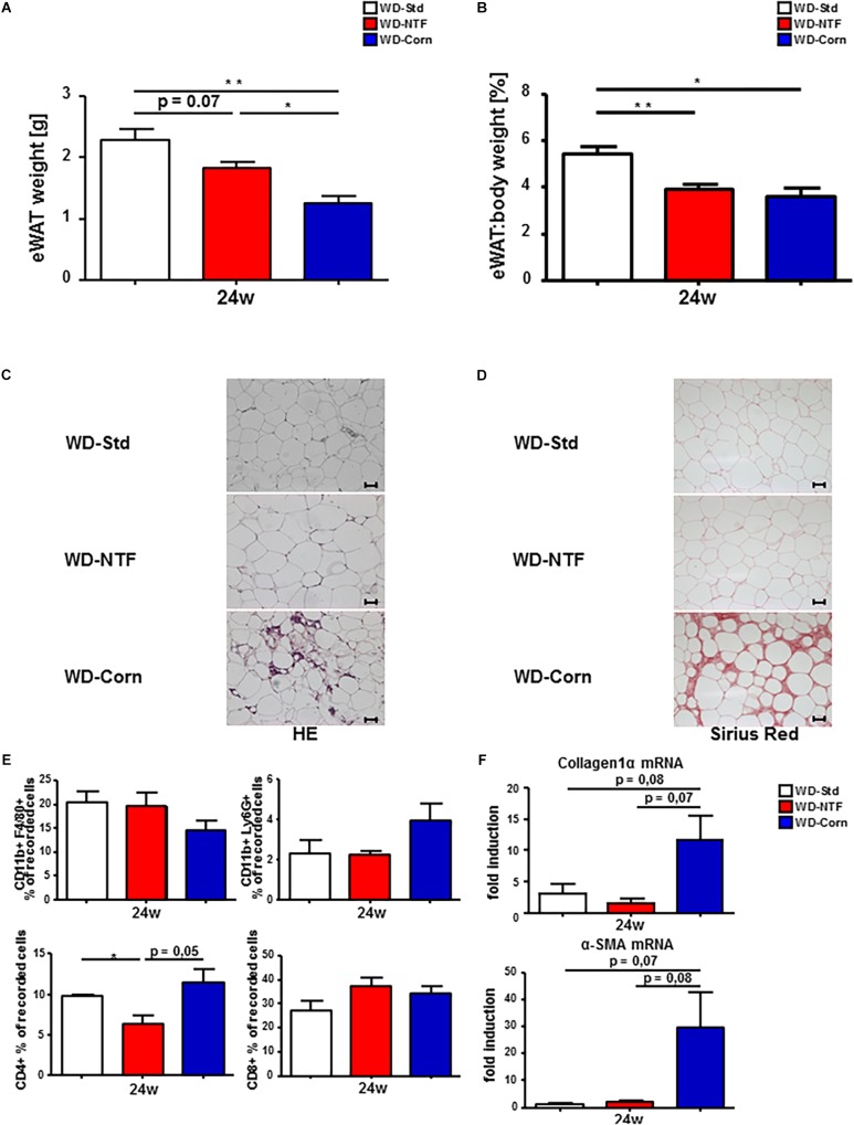
FIGURE 6.
More pronounced fibrosis progression in eWAT of mice treated with WD containing corn oil. (A) Total eWAT weight and (B) eWAT:body weight ratio drops in WT animals fed a WD-NTF compared to standard WD-Std and WD-Corn after 24 weeks treatment (n = 4) (∗p < 0.05, ∗∗ p < 0.01). (C) Representative H&E stained eWAT sections of WT animals after 24 weeks treatment with different Western style diets (WD-Std, WD-NTF, WD-Corn). (D) Representative images of Sirius Red stained eWAT sections from WD fed WT animals (WD-Std, WD-NTF, WD-Corn). (E) Flow Cytometric analysis of eWAT infiltrating immune cells. Cells were gated via FSC/SSC, duplets were excluded. Live/CD45+, CD11b+/F4/80+ (regarded as macrophages) or CD11b+/Ly6G+ (regarded as neutrophils), CD4+ or CD8+ (n = 4) (∗p < 0.05). (F) mRNA expression levels of Collagen1α and α-SMA in whole eWAT homogenates. WT mice were fed WD (WD-Std, WD-NTF, WD-Corn) for 24 weeks. mRNA expression was analyzed via real-time PCR. 4 animals per group were included.

