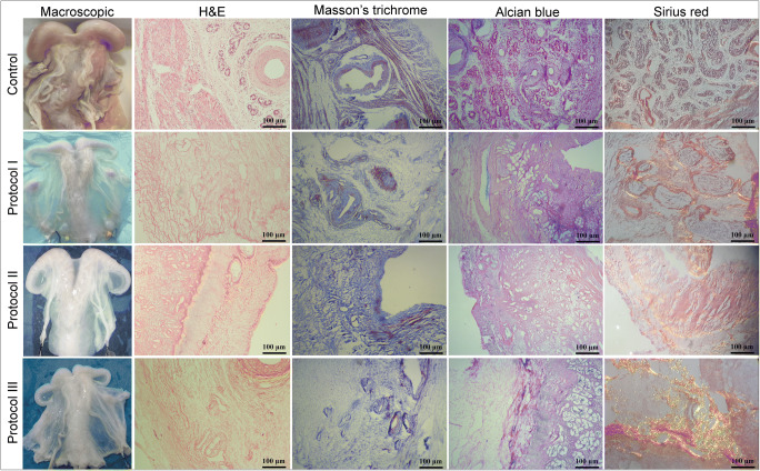
Fig. 1.
Macroscopic evaluation: The whitening and transparency of the organs were considered as a macroscopic sign of organ decellularization. H&E staining: The absence of nuclear components is observed in all specimens which is suggestive of tissue decellularization; ECM structure is maintained, and endometrium and myometrium could be detected in all three groups. Masson’s trichrome staining: The collagen fibers in P2 and P3 samples do not demonstrate considerable differences comparing with the native tissue; the absence of smooth muscle cells could be seen in all of the acellular specimens. Alcian blue staining: GAG proteins demonstrate prominent preservation after the decellularization in all three groups, and no considerable difference could be seen comparing with the native samples. Sirius red staining: Collagen and elastin fibers show notable preservation in all three layers of P2 and P3 samples

