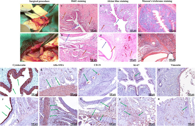
Fig. 6.
Scaffold implantation and histological and immunohistochemical examination. The surgical procedure comprises (A and B) implantation of 10 × 5 mm2 segments of protocol III scaffolds into the right uterine horns of eight female rats using interrupted 5-0 nylon sutures. H&E staining of the native rat uterus (C) and the grafted scaffolds (D) shows the endothelial layer as the inner epithelial layer in both samples. The myometrium including smooth muscle fibers and collagen fibers is detectable in both samples (blue arrows indicate the endometrium, and brown arrows are used to illustrate the myometrium). Alcian blue staining of the control samples (E) and the implanted tissues (F) confirms the structural maintenance of the ECM and recellularization of the grafted samples (blue arrows indicate the endometrium, and brown arrows are used to illustrate the myometrium). Trichrome staining of the controls (G) and the grafts (H) illustrates muscle fibers with red color and the collagen fibers as blue fibers in all of the specimens which are relatively similar in structure and content. Immunohistochemical evaluation (positive-stained cells are in brown color; green arrows indicate stained cells) for cytokeratin marker in the control (I) and the implanted samples (J) shows the endometrium as intact inner epithelial layer in both groups. Alfa-SMA staining of natural rat uterus (K) and implanted acellular samples (L) demonstrates a considerable positive reaction in both groups which confirms the presence of smooth muscle cells in the myometrium. CD31 marker staining of the native (M) and grafted (N) tissues show diffused positive reaction in both groups. The grafted samples demonstrate increased positive-stained cells. IHC staining for Ki-67 in native tissue (O) shows a weak positive reaction, and the implanted scaffolds (P) a prominently higher positive reaction for Ki-67 marker both in the endometrium and the myometrium. IHC staining for vimentin illustrated the diffuse fibro-connective tissue in the myometrium of the native (Q) and grafted (R) specimens with no considerable difference

