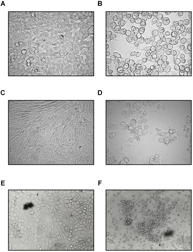
FIGURE 1.

Cytopathic effect of the feline adenovirus isolate in permissive cells as compared to uninfected cells on day 5 postinfection (400×). Uninfected (A) and infected (B) HeLa cells, uninfected (C) and infected CRFK (D) cells, uninfected (E), and infected (F) PD-5 cells. Infected cells form grape-like clusters.
