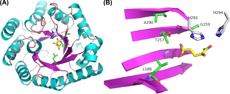
Figure 3. Homology model of MAM1 from A. thaliana.
(A) The model of MAM1 was based on the crystal structure of N. meningitis IPMS (PDB-ID 3RMJ). β sheets (purple) and α helixes (turquoise) comprise the (β/α)8 catalytic barrel characteristic of IPMS/MAM family. The binding of IPMS substrate is shown in yellow. Green residues represent the five residues in MAM1 predicted to be within 8 Å of the substrate. Grey residues represent two His residues conserved in the IPMS/MAM family. (B) Simplified presentation of the residues in MAM1, which is predicted to be in close proximity of the substrate. Colours as in (A).

