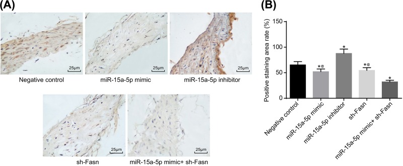
Figure 4. CRP expression by immunohistochemistry.
Tail veins of STZ-induced model rats (n=30 and 5 mice died or were not successfully modeled during STZ induction) were injected with liposomes containing different sequences (miR-15a-5p mimic, miR-15a-5p inhibitor or FASN shRNA) three times a week for 4 weeks. After 4 weeks, the thoracic aortas of each group rats (n=6 for each group) was isolated and used for making paraffin slices and immunohistochemical staining. (A) Staining image from immunohistochemistry of CRP (400×); (B) area percentage of positive CRP. Data shown are mean ± SD, experiments had been repeated three times, *, P<0.05 compared with NC group; @, P<0.05 compared with miR-15a-5p mimic + sh-FASN group.

