Figure 3.
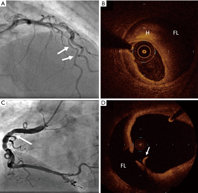
Optical coherence tomography confirmation of SCAD. (A) Angiographic appearance of Type 3 SCAD of the left anterior descending artery (arrows), confirmed with (B) optical coherence tomography imaging, showing intramural haematoma (H) and false lumen (FL). (C) Angiogram image of right coronary artery in patient who presented with acute coronary syndrome showing beaded appearance (arrow) and (D) accompanying optical coherence tomography image showing an intimal flap (arrow) and false lumen (FL). SCAD, spontaneous coronary artery dissection.

