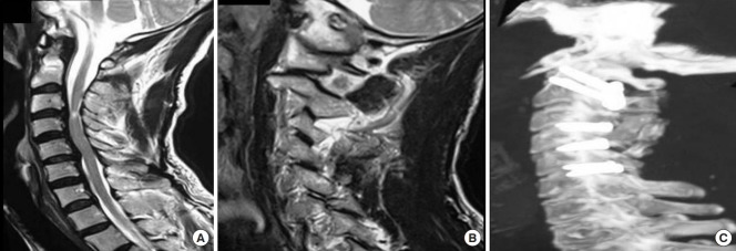
Fig. 5.
Images of a 52-year-old male patient. (A) T2-weighted magnetic resonance imaging (MRI) showing multisegmental spinal degeneration with evidences of cord compression and cord signal alteration. There is no neural compression in the region of craniovertebral junction. (B) MRI cut through the facets of atlas and axis showing type 2 atlantoaxial facetal instability. (C) Postoperative X-ray showing multisegmental fixation that includes atlantoaxial joint fixation.

