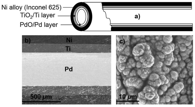
Figure 3.
Images of the catalytic tubular reactor: (a) Schematic presentation of the tubular reactor; (b) Energy-dispersive X-ray spectroscopy (EDX) mapping of the longitudinal section of the Ni alloy (Inconel 625) tube with the TiO2/Ti secondary layer coated with the thin Pd layer; (c) Scanning electron microscopy (SEM) image of deposited Pd [180].

