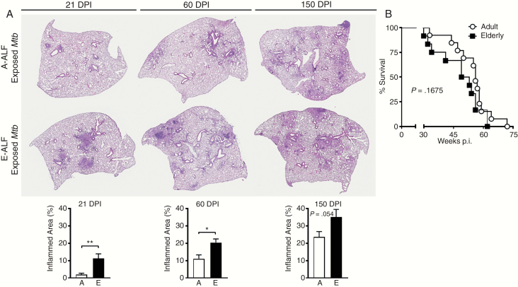
Figure 6.
A, Elderly alveolar lining fluid (E-ALF, “E”)–exposed Mycobacterium tuberculosis (Mtb) demonstrates increased Mtb-induced immunopathology in the lung. C57BL/6J mice were infected with a low-dose aerosol of Mtb strain Erdman previously exposed to adult alveolar lining fluid (A-ALF, “A”) or E-ALF and washed. At 21, 60, and 150 days postinfection (DPI), mice were euthanized and lungs fixed in 10% neutral buffered formalin, embedded in paraffin, sectioned, and stained with hematoxylin and eosin to visualize tissue morphology. Areas of cell aggregation and infiltration (inflammation) were quantified using Aperio Imagescope by calculating the area of inflammatory foci (ie, involvement) divided by the total area of the lung. Quantification for 21, 60, and 150 DPI are shown. Representative images at a final magnification of ×20. Pooled results from n = 2 with 4–5 mice/group, using 2 A-ALFs and 2-E-ALFs, a set of different A-ALF vs E-ALF in each experiment, mean ± standard error of the mean; Student t test. *P < .05; **P < .005. B, Survival was monitored across a period of 80 weeks postinfection (p.i.). Mice were euthanized when they met the exclusion criteria documented in animal care and use protocols, and the date was documented. Pooled experiment from n = 2 with 10–20 mice; using 2 A-ALFs and 2-E-ALFs, a set of different A-ALF vs E-ALF in each experiment. Log-rank test.

