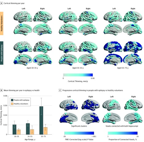
Figure 1. Progressive Epilepsy-Associated Cortical Thinning Compared With Thinning Associated With Normal Aging.
A, Annualized rate of cortical thinning in healthy volunteers (top) and people with epilepsy (bottom), stratified to 3 age groups (18 to <35, 35 to <55, and 55 to <75 years). B, Comparison of annualized cortical thinning rates between people with epilepsy and controls (vertical lines indicate SEM). C, Vertexwise statistical comparison of progressive cortical thinning in people with epilepsy vs healthy volunteers using linear mixed-effects models. Hemispheric surface templates are displaying a map of significant clusters after random field theory correction for multiple comparisons (left). Structural connectivity with left and right hippocampi in 10 healthy volunteers is presented as the regional proportion of connected voxels (right). FWE indicates familywise error.

