Fig. 3.
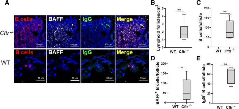
LFs, B cells, BAFF+ and IgG+ B cells were increased in uninfected Cftr −/− mice. a Representative confocal images of triple-color immunofluorescence staining of representative pulmonary LFs from uninfected Cftr −/− mice (top two rows) and wild type (WT) controls (bottom row) from cohort 1. B cells are identified by staining for CD45R and red fluorophore. BAFF positive cells have a grey color. IgG positive B cells were identified by staining with green fluorophore. 4′6-diamidino-2-phenylindole (blue) was used to counterstain the nuclei. The final panel in each row is a merged file of CD45R, BAFF and IgG staining. In the Cftr −/− mice images (top row), the magnification is 60X. In the wild type control images (bottom row), the magnification is 100X. The images shown are representative of LFs in Cftr −/− mice (n = 6) and WT controls (n = 7). Cftr −/− and WT lung b Lymphoid follicles, c B cells, d BAFF+ B cells and e IgG+ B cells were quantified. Mann-Whitney U test was used to perform the statistical analysis (b-e). Box plots show the median values and 25th and 75th percentiles, and error bars show the 10th and 90th percentiles. *p ≤ 0.05, **p ≤ 0.001, Cftr −/− mice versus WT

