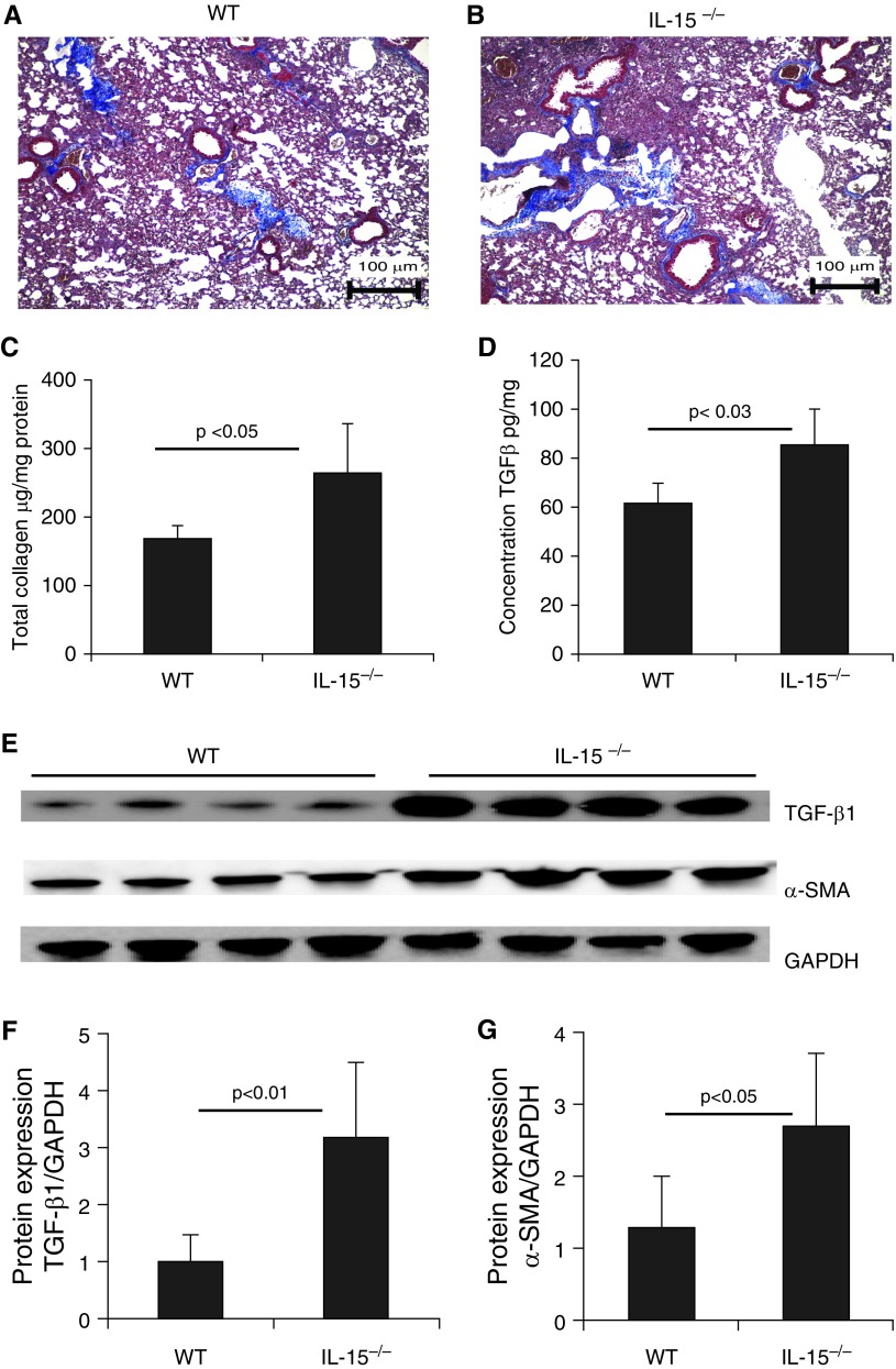
Figure 1.
Analysis of baseline tissue remodeling in endogenous IL-15 gene–deficient (IL-15−/−) mice. (A and B) Baseline collagen deposition was examined using Masson’s trichrome staining of lung tissue sections from 20-weeks-old naive wild-type (WT) and (C and D) IL-15−/− mice. (C) Total lung collagen was assessed using Sircol reagent, and (D) transforming growth factor β1 (TGF-β1) levels were assessed by ELISA. (E–G) Western blot analysis showed enhanced expression of TGF-β1 and α-SMA in IL-15−/− mice (E), which was confirmed by performing GAPDH-normalized densitometry (F and G). Data are expressed as mean ± SD, n = 8 mice/group in each group, except for the Western blot analysis. Scale bars: 100 μm. SMA = smooth muscle actin.

