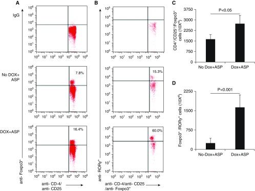
Figure 6.
Analysis of T regulatory (Treg) cells in DOX-regulated CC10–IL-15 transgenic mice after Aspergillus challenge. (A and B) Treg cells in the mediastinal lymph nodes of Aspergillus-challenged DOX- and non-DOX–exposed CC10–IL-15 overexpressing mice were examined by flow cytometry using anti-CD45-FITC, anti-CD4-PerCP, anti-RORγ-PE, anti-CD25-PE-Cy7, and anti-Foxp3-APC along with matched IgG antibodies. (C) The average percentage of CD4+CD25+Foxp3+ and (D) RORγ+CD4+CD25+Foxp3+ Treg cells, and their absolute number of cells from three independent experiments in mediastinal lymph nodes of mice. Data are expressed as mean ± SD.

